 Figure 1a, b.
Full A. fundyense cyst and empty A. fundyense cyst side by side
(empty is above full) in
epifluorescent and transmitted light. Primulin stained. 200x magnification.
Figure 1a, b.
Full A. fundyense cyst and empty A. fundyense cyst side by side
(empty is above full) in
epifluorescent and transmitted light. Primulin stained. 200x magnification.University of Maine,
School of Marine Sciences, Orono, Maine.
This is a working page for the ECOHAB-Gulf of Maine project participants, and is intended to facilitate dialogue between concerned parties.
This page contains images of Alexandrium sp. cysts as well as some unidentified
dinoflagellate cysts and other similar objects. The pictures were taken with a
Nikon Optiphot-2 Epifluorescence microscope. Magnification of individual images is
given in each caption. Please do not link this page to any other site.
No image may be copied, downloaded, reproduced, or used in any other manner, etc. without permission from Sarah Kirn.
(207)581-4348, 5741 Libby Hall, University of Maine, Orono, ME 04469-5741
NOTE: There exists some controversy regarding whether the dominant Alexandrium species in the Gulf of Maine is A. tamarense or A. fundyense. I am unfamiliar with the distinction between A. tamarense and A. fundyense; purely for reasons of clarity when distinguishing between A. ostenfeldii (which has been observed in low densities in the Gulf) and the dominant Alexandrium species in the Gulf, I refer to the latter as A. fundyense.
Alexandrium fundyense cysts
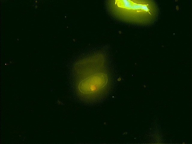
Figure 1a, b.
Full A. fundyense cyst and empty A. fundyense cyst side by side
(empty is above full) in
epifluorescent and transmitted light. Primulin stained. 200x magnification.
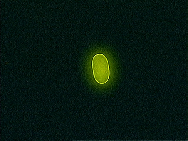
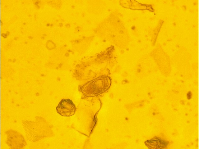
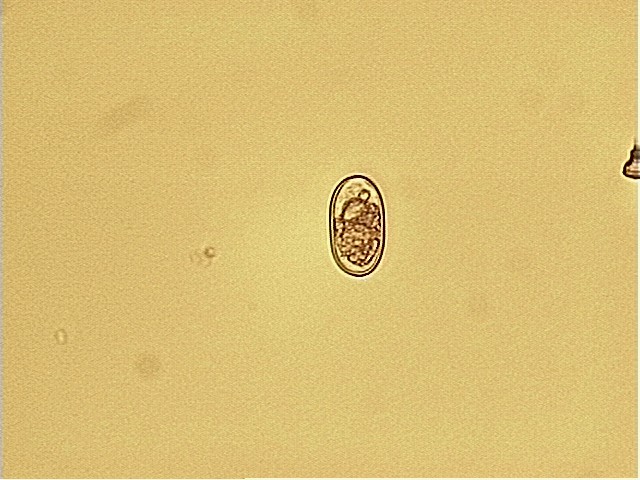 Figure 2a, b. Possible A. fundyense cyst - notice lack of structure
in epifluorescent micrograph compared to micrograph below. Primulin
stained. Epifluorescent and transmitted light, 200x magnification.
Figure 2a, b. Possible A. fundyense cyst - notice lack of structure
in epifluorescent micrograph compared to micrograph below. Primulin
stained. Epifluorescent and transmitted light, 200x magnification.
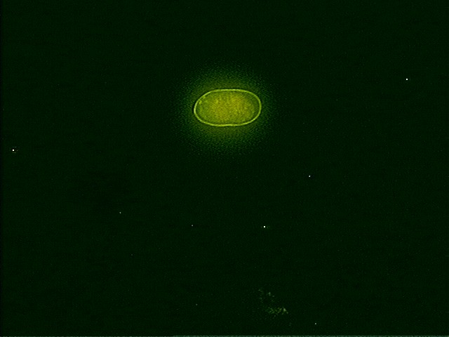
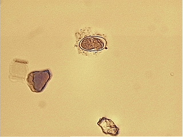 Figure 3a, b.
A. fundyense cyst in epifluorescent and transmitted light. Primulin
stained. 200x magnification.
Figure 3a, b.
A. fundyense cyst in epifluorescent and transmitted light. Primulin
stained. 200x magnification.
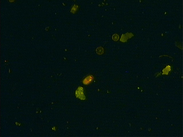
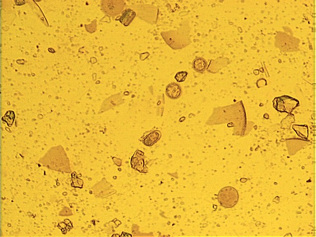 Figure 4a, b.
A. fundyense cyst exhibiting chlorophyll excitation (looks red under epifluorescent light).
Primulin stained. Epifluorescent and transmitted light, 100x magnification.
Figure 4a, b.
A. fundyense cyst exhibiting chlorophyll excitation (looks red under epifluorescent light).
Primulin stained. Epifluorescent and transmitted light, 100x magnification.
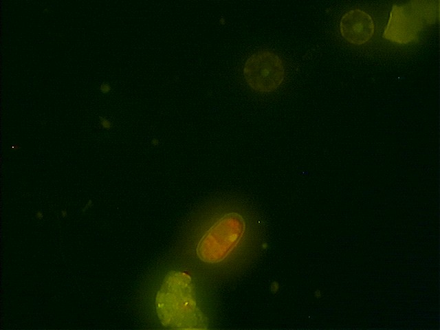
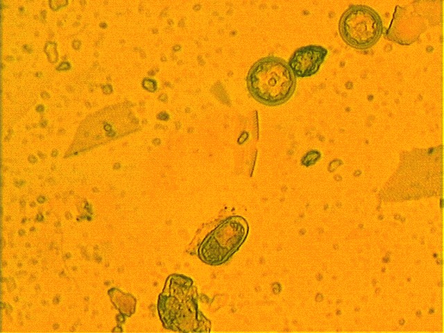 Figure 4c, d. Same cyst as
above under 200x magnification. Primulin stained. Epifluorescent and transmitted light.
Figure 4c, d. Same cyst as
above under 200x magnification. Primulin stained. Epifluorescent and transmitted light.
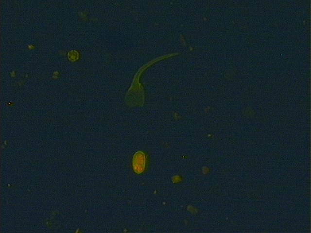
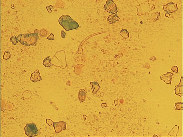 Figure 5a, b.
A. fundyense cyst. Primulin stained. Epifluorescent and transmitted light,
100x magnification.
Figure 5a, b.
A. fundyense cyst. Primulin stained. Epifluorescent and transmitted light,
100x magnification.
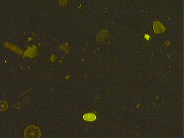
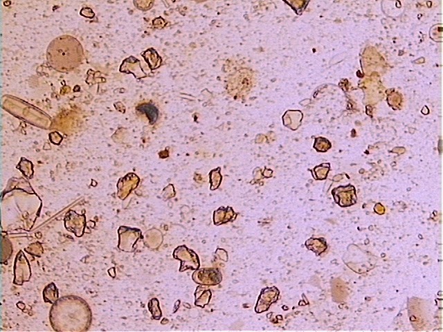 Figure 6a, b. Empty A. fundyense cyst.
Primulin stained. Epifluorescent and transmitted light, 100x magnification.
Figure 6a, b. Empty A. fundyense cyst.
Primulin stained. Epifluorescent and transmitted light, 100x magnification.
Odd-looking Alexandrium cysts
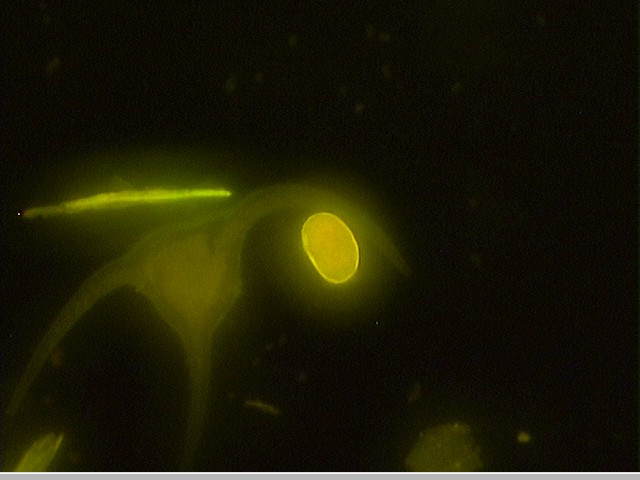
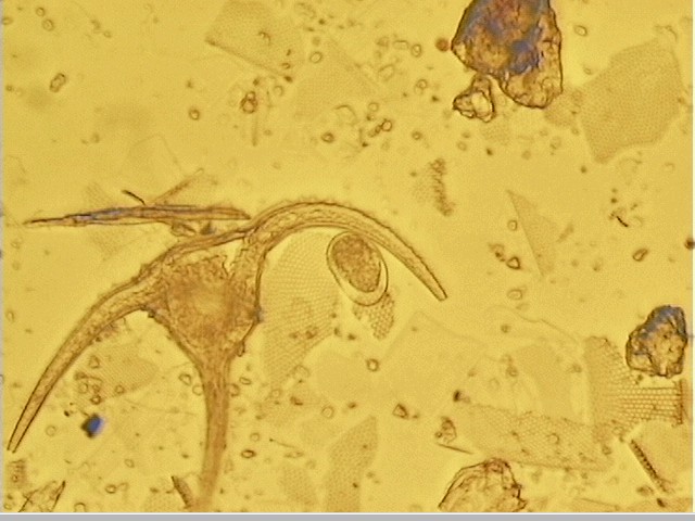 Figure 7a, b. Cyst with no internal definition under
epifluorescent light, but normal-looking in transmitted light. Primulin
stained, epifluorescent and transmitted light, 200x magnification.
Figure 7a, b. Cyst with no internal definition under
epifluorescent light, but normal-looking in transmitted light. Primulin
stained, epifluorescent and transmitted light, 200x magnification.
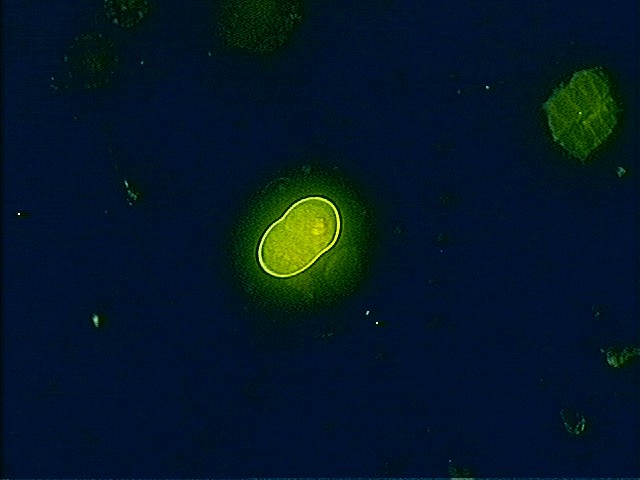
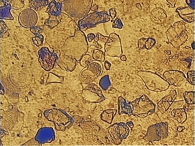 Figure 8 a, b. Same as above, second example. Primulin stained,
epifluorescent and
transmitted light, 200x magnification.
Figure 8 a, b. Same as above, second example. Primulin stained,
epifluorescent and
transmitted light, 200x magnification.
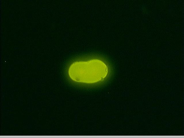
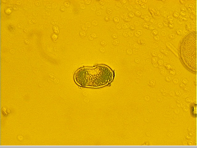 Figure 9 a, b. Opaque cyst, primulin stained, 64 um long by 32 um wide. Epifluorescent
and transmitted light, 200x magnification.
Figure 9 a, b. Opaque cyst, primulin stained, 64 um long by 32 um wide. Epifluorescent
and transmitted light, 200x magnification.
Unidentified - possibly A. ostenfeldii cysts, some other dinoflagellate cyst, or copepod eggs
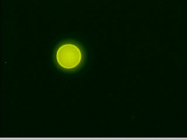
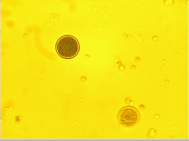 Figure 10 a, b. Primulin stained cell, 40 um diameter. Epifluorescent and
transmitted light, 200x magnification.
Figure 10 a, b. Primulin stained cell, 40 um diameter. Epifluorescent and
transmitted light, 200x magnification.
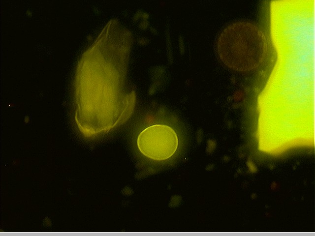
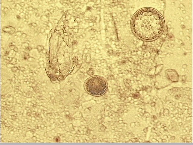 Figure 11 a, b. Primulin stained cell, 40 x 32 um. Epifluorescent and
transmitted light, 200x magnification.
Figure 11 a, b. Primulin stained cell, 40 x 32 um. Epifluorescent and
transmitted light, 200x magnification.
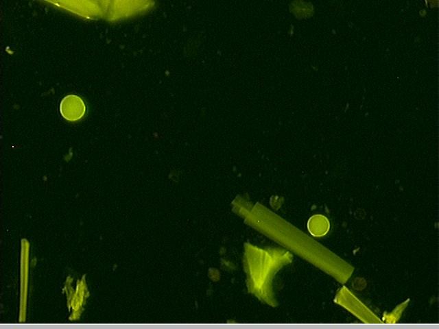
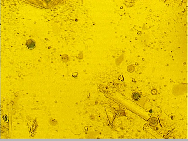 Figure 12 a, b. Two round, double-walled cells stained with
primulin. Cell in upper left is 40 um across (see enlarged view in 12 c,
d), cell in lower right is 32 um across (see enlarged view in 12 e, f)..
Figure 12 a, b. Two round, double-walled cells stained with
primulin. Cell in upper left is 40 um across (see enlarged view in 12 c,
d), cell in lower right is 32 um across (see enlarged view in 12 e, f)..
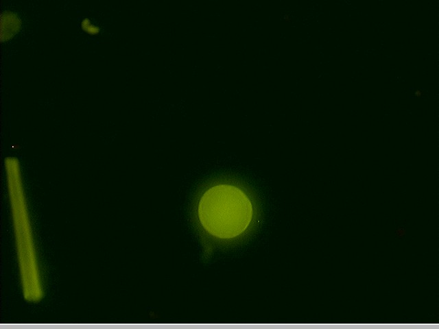
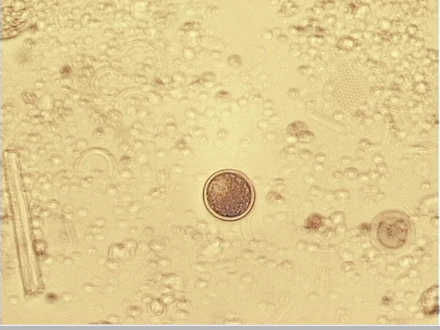 Figure 12 c,d.
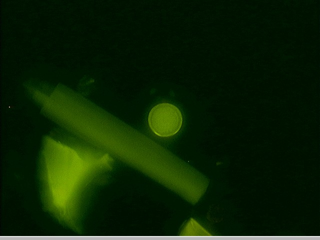
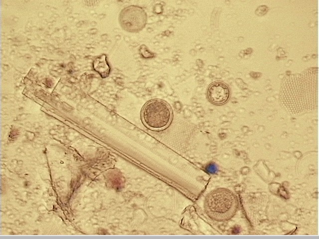 Figure 12 e, f. 40 um cell from 12 a, b. 200x
magnification.
32 um cell from 12 a,b. 200x magnification.
Figure 12 e, f. 40 um cell from 12 a, b. 200x
magnification.
32 um cell from 12 a,b. 200x magnification.
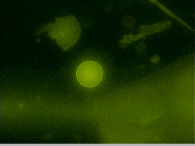
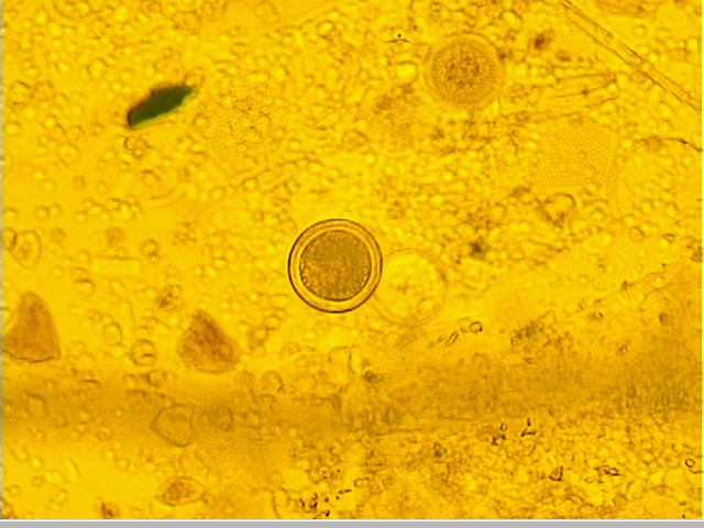 Figure 13 a, b. Primulin stained cell, approximately 48 um across. Note that internal contents
have pulled away from cell wall. Epifluorescent and
transmitted light, 200x magnification.
Figure 13 a, b. Primulin stained cell, approximately 48 um across. Note that internal contents
have pulled away from cell wall. Epifluorescent and
transmitted light, 200x magnification.
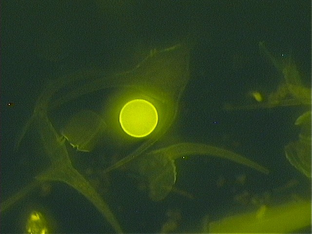
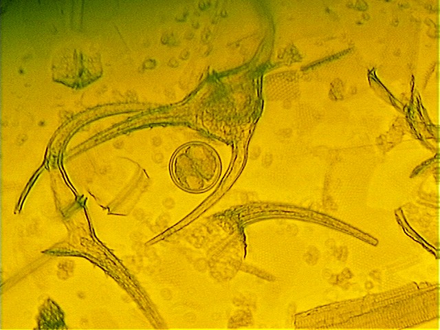 Figure 14 a, b. Primulin stained cell, 38 um across. Note that internal contents
appear to be in two bundles. Epifluorescent and
transmitted light, 200x magnification.
Figure 14 a, b. Primulin stained cell, 38 um across. Note that internal contents
appear to be in two bundles. Epifluorescent and
transmitted light, 200x magnification.
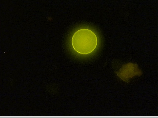
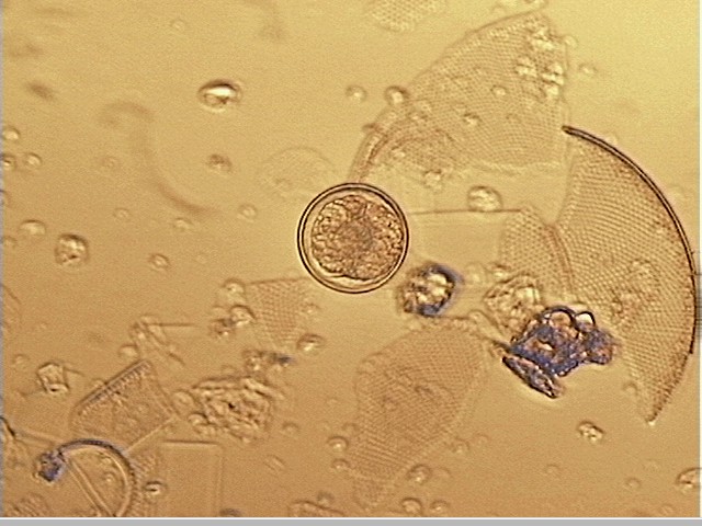 Figure 15 a, b. Primulin stained cell, 48 um across. Epifluorescent and
transmitted light, 200x magnification.
Figure 15 a, b. Primulin stained cell, 48 um across. Epifluorescent and
transmitted light, 200x magnification.
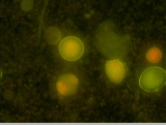
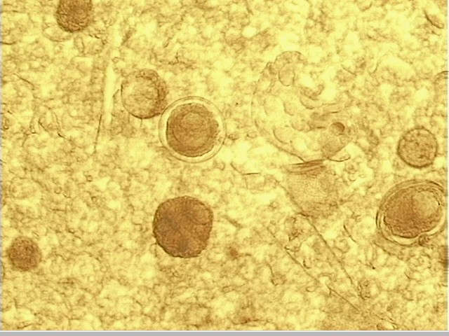 Figure 16 a, b. Primulin stained cell, 50 um across. Epifluorescent
and transmitted light, 200x magnification.
Figure 16 a, b. Primulin stained cell, 50 um across. Epifluorescent
and transmitted light, 200x magnification.
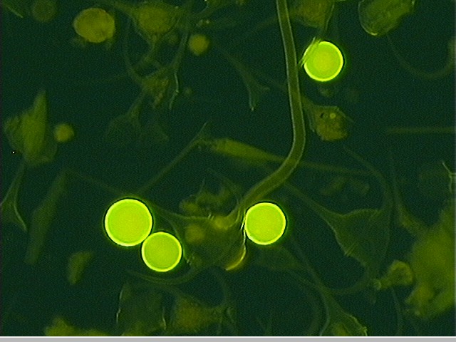
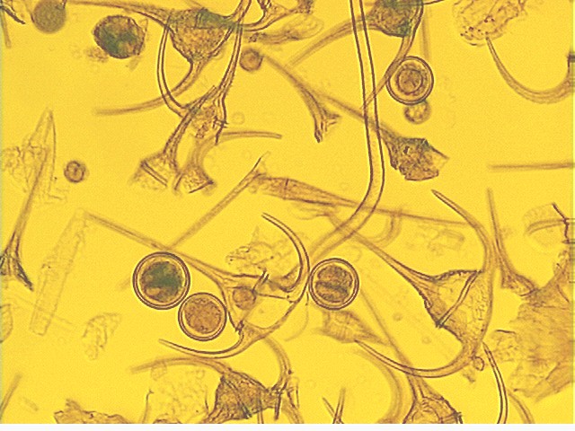 Figure 17 a, b. Note especially the difference in appearance of the
internal material in each cell. Primulin stained. Epifluorescent and
transmitted light, 100x magnification.
Figure 17 a, b. Note especially the difference in appearance of the
internal material in each cell. Primulin stained. Epifluorescent and
transmitted light, 100x magnification.
Micrographs of live samples of same unidentified round cells with inclusion bodies as seen above
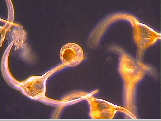 Figure 18.
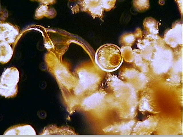
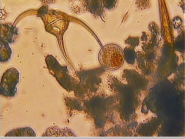 Figure 19 a, b.
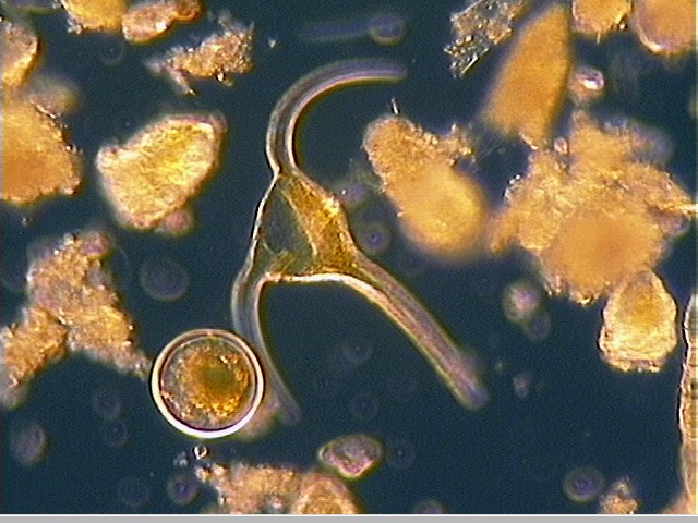
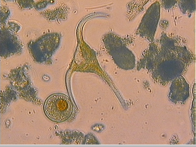 Figure 20 a, b.