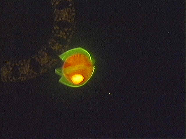
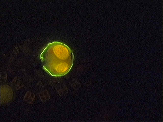
 Image 1. St. 67 (2m) of cruise CH0900
April/May ECOHAB; large cell approx. 54um theca breaking, note dense,
yellow,vacuolar material viewed at 200X magnification.
Image 1. St. 67 (2m) of cruise CH0900
April/May ECOHAB; large cell approx. 54um theca breaking, note dense,
yellow,vacuolar material viewed at 200X magnification.University of Maine School of Marine Sciences ECOHAB-GOM Project 2000
The following are images of Alexandrium sp. All were taken on a Nikon Optiphot-2 Epifluorescence microscope and CCD camera. Please do not link this page to any other site. Also, please obtain permission from Abigail Deitz: contact abigail.deitz@umit.maine.edu or (207) 581-4314, 5741 Libby Hall, University of Maine, Orono, ME 04469-574, before using images outside the ECOHAB-GOM working group.
To reach Sarah Kirn's water column cyst observations, complete with pictures, use this link: /Web-Pictures/Web-Pictures.htm
Image 1. St. 67 (2m) of cruise CH0900
April/May ECOHAB; large cell approx. 54um theca breaking, note dense,
yellow,vacuolar material viewed at 200X magnification.
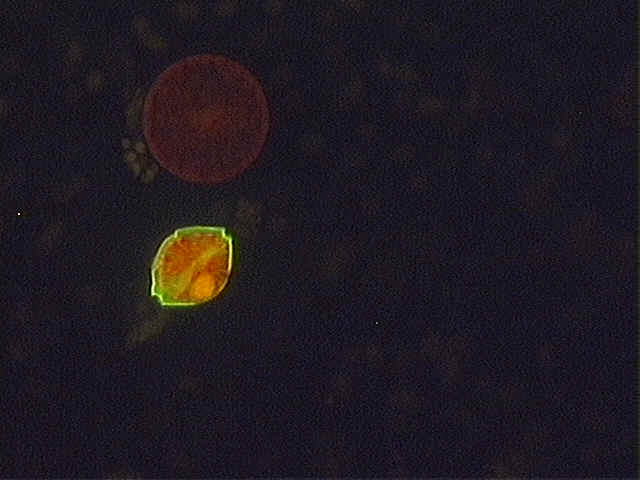 Image 2. St. 67 (2m)cruise CH0900;
elongated/compressed cell with positive reaction to fluoroscein viewed at 200X
Image 2. St. 67 (2m)cruise CH0900;
elongated/compressed cell with positive reaction to fluoroscein viewed at 200X
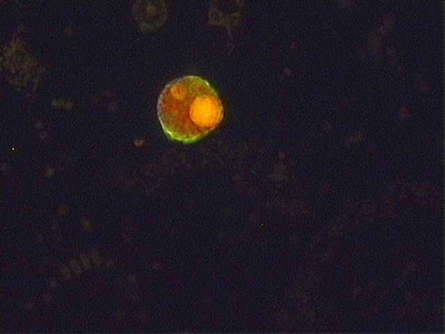 Image 3. St. 67 (2m) CH0900; 53um
cell with dense, golden-colored inclusions, with a positive reaction but not strong, 200X
Image 3. St. 67 (2m) CH0900; 53um
cell with dense, golden-colored inclusions, with a positive reaction but not strong, 200X
 Image 4. St.
67 (2m) CH0900; similar cell to Image 3. but negative reaction to fluoroscein ,200X
Image 4. St.
67 (2m) CH0900; similar cell to Image 3. but negative reaction to fluoroscein ,200X
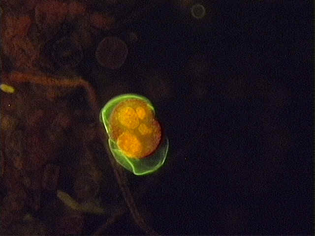
 Image
5. St. 67 (2m) CH0900; large cell, 54um, breaking viewed at, 200X
Image
5. St. 67 (2m) CH0900; large cell, 54um, breaking viewed at, 200X
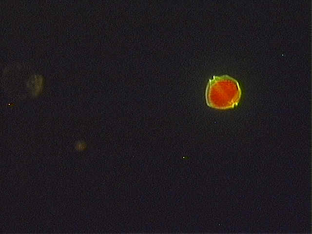 Image 6. St. 67 (2m)CH0900; vegetative cell approx. 30-32um
viewed at 200X, compare to size of larger cells, these are the "normal" cells
(in size and morphology)we look for with this staining procedure.
Image 6. St. 67 (2m)CH0900; vegetative cell approx. 30-32um
viewed at 200X, compare to size of larger cells, these are the "normal" cells
(in size and morphology)we look for with this staining procedure.
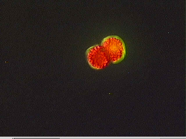 Image 7. St. 45 (2m) CH0900; caught in the act of cell
division
Image 7. St. 45 (2m) CH0900; caught in the act of cell
division
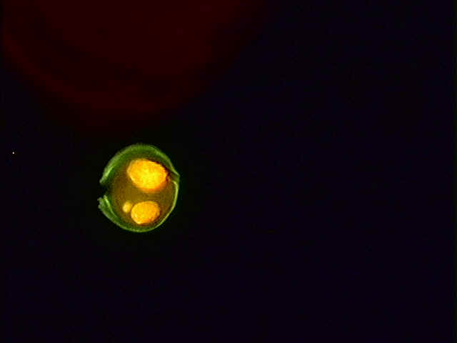 Image 8. St. 8 (30m) collected on
cruise END336 first week of April 2000, again a 54um cell full of dense, golden
inclusions, note the lack of chlorophyll fluorescence. Is this an heterotrophic A.
ostenfeldii? 200X
Image 8. St. 8 (30m) collected on
cruise END336 first week of April 2000, again a 54um cell full of dense, golden
inclusions, note the lack of chlorophyll fluorescence. Is this an heterotrophic A.
ostenfeldii? 200X
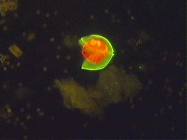
 Image
9. St. 8 (30m) END 336;viewed at 200X one of these large cells
is breaking apart. Note that theca breaking away is strongly stained with fluorescence but
leaving behind just the red and gold colors of chlorophyll and inclusions....hmmm...would
it have looked like Image 4 had the theca separated completely?
Image
9. St. 8 (30m) END 336;viewed at 200X one of these large cells
is breaking apart. Note that theca breaking away is strongly stained with fluorescence but
leaving behind just the red and gold colors of chlorophyll and inclusions....hmmm...would
it have looked like Image 4 had the theca separated completely?
 Image
10a. St. 9 (2m) END 336; 200X; large(54um) reactive cell, low chlorophyll
fluorescence
Image
10a. St. 9 (2m) END 336; 200X; large(54um) reactive cell, low chlorophyll
fluorescence
 Image 10b. Same cell in 10a. under tungsten light
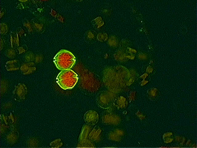 Image 11. A duplet from St. 104, June 2000, 2m.
There were a number of duplets in this particular sample and some in surrounding stations
as well. Viewed at 200X.
Image 11. A duplet from St. 104, June 2000, 2m.
There were a number of duplets in this particular sample and some in surrounding stations
as well. Viewed at 200X.
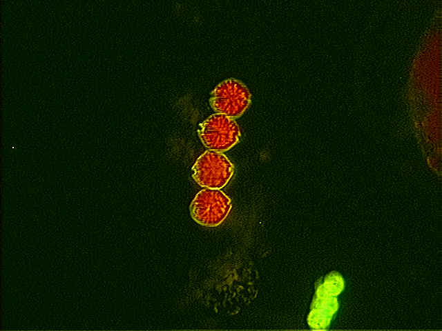 Image12. Chain of four cells seen at St. 23 from the
June 2000 cruise. The occurrence of quadruplet chains in this year's samples has
been frequent.
Image12. Chain of four cells seen at St. 23 from the
June 2000 cruise. The occurrence of quadruplet chains in this year's samples has
been frequent.
 Image 13. Chain of three cells, note
gradation in size, larger cell to left, smaller cell on right. This particular
picture is difficult to see with regards to fluorescence due to photobleaching. Two Ceratium
cells are seen to the far left of the chain.
Image 13. Chain of three cells, note
gradation in size, larger cell to left, smaller cell on right. This particular
picture is difficult to see with regards to fluorescence due to photobleaching. Two Ceratium
cells are seen to the far left of the chain.
 Image
14. Alexandrium ostenfeldii as seen by electron microscopy.
Sample taken from St. 342 collected on cruise CH09, April/May 2000. Approximate
location at 43° 43.51" and 69° 54.44". Note shape of primary apical plate with
large pore, distinctive of A. ostenfeldii as described by Tomas in Identifying
Marine Phytoplankton pg. 496 and 499.
Image
14. Alexandrium ostenfeldii as seen by electron microscopy.
Sample taken from St. 342 collected on cruise CH09, April/May 2000. Approximate
location at 43° 43.51" and 69° 54.44". Note shape of primary apical plate with
large pore, distinctive of A. ostenfeldii as described by Tomas in Identifying
Marine Phytoplankton pg. 496 and 499.
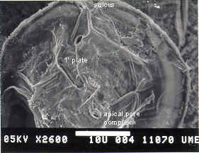 Image 15. Apical view of both pore complex and first apical
plate labelled.
Image 15. Apical view of both pore complex and first apical
plate labelled.
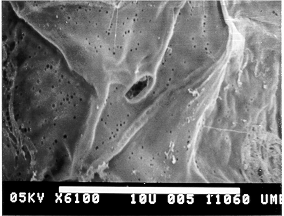 Image
16. Close up view of this primary apical plate.
Image
16. Close up view of this primary apical plate.
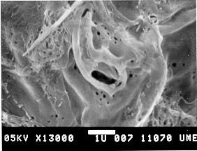 Image
17. Close up view of apical pore complex.
Image
17. Close up view of apical pore complex.
. Image 18. Calcofluor stain used to highlight apical plate.
Magnification at 400X. This cell located in sample from St. 346 of the CH09
April/May 2000 cruise.
Image 18. Calcofluor stain used to highlight apical plate.
Magnification at 400X. This cell located in sample from St. 346 of the CH09
April/May 2000 cruise.
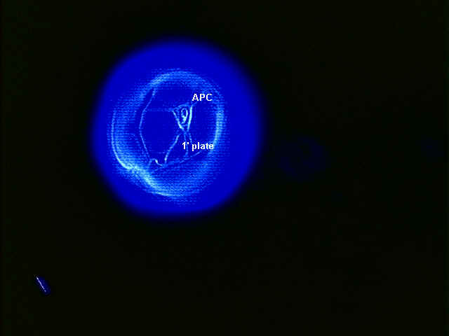 Image 19. Apical view of a calcofluor stained cell.
St. 346 April/May cruise. Apical pore complex (APC) and primary apical plate (1' plate)
are labelled.
Image 19. Apical view of a calcofluor stained cell.
St. 346 April/May cruise. Apical pore complex (APC) and primary apical plate (1' plate)
are labelled.